Ciril Bohak
Žiga Lesar
Manca Žerovnik Mekuč
Eva Boneš
Matija Marolt
University of Ljubljana, Faculty of Computer and Information Science, Laboratory for Computer Graphics and Multimedia
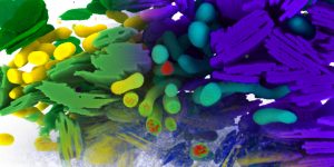
Intracellular compartments acquired using the FIB-SEM technique. In the top right is the Ground Truth (GT) data, in the top left the automatically segmented (SEG) data, and in the bottom the raw acquired image. Blending of all three images showing how well does the automatic segmentation method work for mitochondria (GT – blue, SEG – yellow) and fusiform vesicles (GT – violet, SEG – green).


