Lara Tomholt, Wyss Institute for Biologically Inspired Engineering, Harvard University, Cambridge and Harvard Graduate School of Design, Harvard University, Cambridge
James C. Weaver, School of Engineering and Applied Sciences, Wyss Institute for Biologically Inspired Engineering, Harvard University, Cambridge
Mason N. Dean; Max Planck Institute of Colloids and Interfaces, Department of Biomaterials, Research Campus Golm, Potsdam, Germany
Hans-Christian Hege; Zuse Institute Berlin, Department of Visual and Data-Centric Computing, Berlin, Germany
Daniel Baum; Zuse Institute Berlin, Department of Visual and Data-Centric Computing, Berlin, Germany
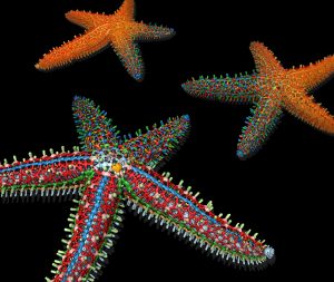
A single starfish (Pisaster giganteus) from the temperate Eastern Pacific was imaged using micro-computed tomography, and its endoskeletal elements (ossicles) were segmented using random-walk distance transform and contour-tree segmentation. The intensity image was first automatically segmented into individual ossicles, which were subsequently classified. The image shows the three main steps of the workflow by blending different renderings.


